The Cornea
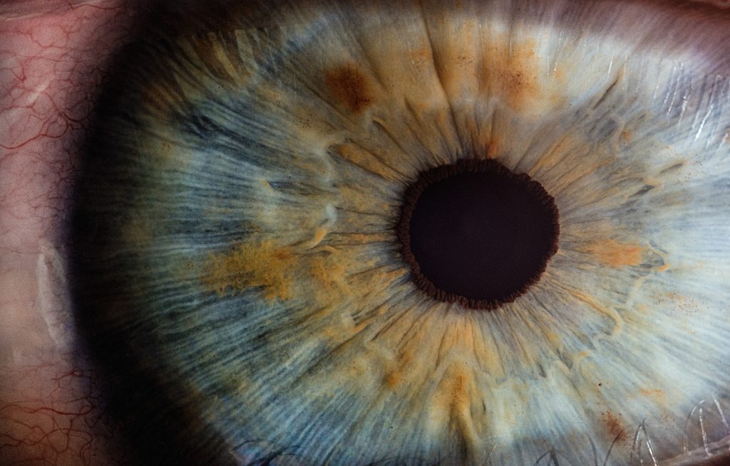
- It’s the outer most layer of the eye, domed shaped, transparent tissue that covers the eyes.
- It has about three quarters of the optical power of the eye
- It is inserted into the sclera at the limbus, about 540 micrometer thick centrally
Facts: Did u know!!! the cornea is the mostly densely innervated tissues in the body!!
- Supplied by the first branch of trigeminal nerve
- It has no blood vessels although it is alive, it receives all the nutrients from the aqueous humor (posteriorly) and tears (anteriorly).
Cornea transparency is due to;
- Uniform structure
- A vascularity
- Deturgescence (relative dehydration)
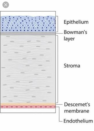
The cornea has five layers ;
- The epithelium; Is stratified squamous and non-keratinizedIt has nerve endings and acts as a barrier of the eye to foreign material eg dust. It absorbs oxygen and nutrients from tears
- Bowman’s membrane It is formed by collagen fibers, when injured, it forms scar as it heals and if the scar will form centrally there will be visual loss
- The stroma contributes to about 90% of the corneal thickness. It’s mainly water and collagens, collagens are the ones that give the cornea its structure and elasticity. The stoma can scar but cannot regenerate
- Descement’s membrane It has regenerative potentials
- The endothelium maintains corneal deturgescence by pumping excess fluid out of the stoma, failure of the endothelium to do its work can lead to cornea edema Its cells cannot regenerate. There is a loss of cells in this layer as the age increases
Functions of the cornea
- It helps in focusing the light that come into the eye so as it can fall on the retina.
- It’s a physical barrier together with tears and eyelid against the dirty and germs that come into contact with the eye.
- It also protect the eye against the UV light from the sun.
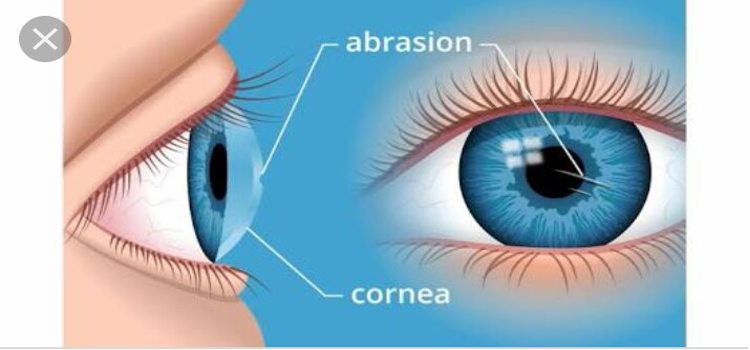
Note: The cornea can be affected by different diseases, keratoconus being among them.
Keratoconus
- It’s a disease characterized by progressive thinning of the cornea and takes a cone shape which results to astigmatism (the cornea cannot focus the light to the retina).
- Most of refraction of light occurs at the air –cornea interface so change in the cornea shape can lead to major impact on vision.
Signs
- Bulging of the lower eyelid on down gazing(munson sign)
- Oil droplets reflex on direct ophthalmoscope
- Scissors reflex on retinoscopy
- Stromal scarring this occurs when descement’s membrane has rupture
Causes
- Some research suggests that it’s due to imbalance of enzymes in the cornea which leads to oxidative damages hence weakening and bulging of the cornea.
- Overexposure to the ultraviolet light,
- Excessive rubbing of the eye,
- Poor fitted contact lens
- Genetic predisposition.
Can glasses correct the keratoconus?
- Glasses can only correct the regular astigmatism but as for keratoconus it is an irregular astigmatism.
- Contact lenses can be used to make the air –cornea interface more spherical so as the light can focus to the eye.
Management options:
- Contact lenses
- Collagens cross linking; it prevent the progression of early form of the disease (riboflavin eye drops which is then exposed to the sun light)
- Corneal graft
- Corneal ring segment implant

Responses