Madura Foot: A Comprehensive Guide
This blog will provide you with a comprehensive guide on Madura foot. It includes all the necessary information on its epidemiology, types, risk factors, clinical features, investigations, differential diagnosis, management, complications, and prognosis.
Introduction:
Madura foot, also known as mycetoma, is a chronic fungal or bacterial infection that affects the subcutaneous tissues of the foot or hand. It can also defined as a chronic granulomatous disease characterized by localized infection of subcutaneous tissues by actinomycetes or fungi. It is a rare disease that is endemic in certain parts of the world, such as India, Africa, and Central and South America. Madura foot is a challenging disease to manage due to its chronic nature and the difficulty in diagnosing it. Was named after a regional district of its discovery, Madurai where it was first described in 1842.
Epidemiology: Madura foot is more prevalent in males than females and typically affects people between the ages of 20 and 40. It is endemic in certain regions where people are more likely to be exposed to the causative agents. The highest incidence of mycetoma is reported in Sudan, followed by India and Mexico.
Types: Madura foot can be classified into two types: eumycetoma (40%), which is caused by fungi, and actinomycetoma, which is caused by bacteria (60%).
Risk Factors: People who are at risk of developing Madura foot include farmers, gardeners, and other individuals who have prolonged exposure to soil and plant material. It is also common in individuals who have a weakened immune system, such as those with HIV/AIDS.
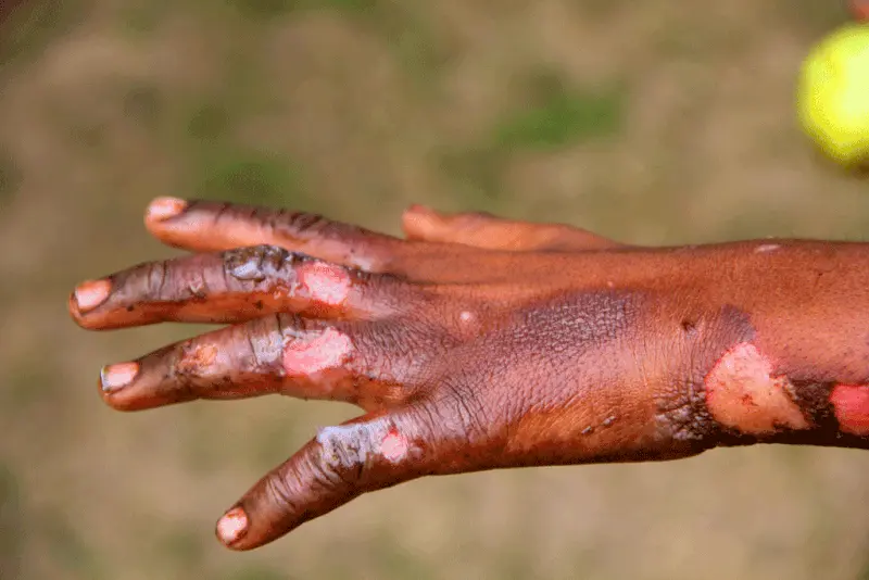
Infection enters through sites of local trauma, e.g. cut or splinter, causing a granulomatous reaction. Spread occurs through skin facial planes and can involve the bone.It most commonly involves the foot but can involve the hands, back or shoulders
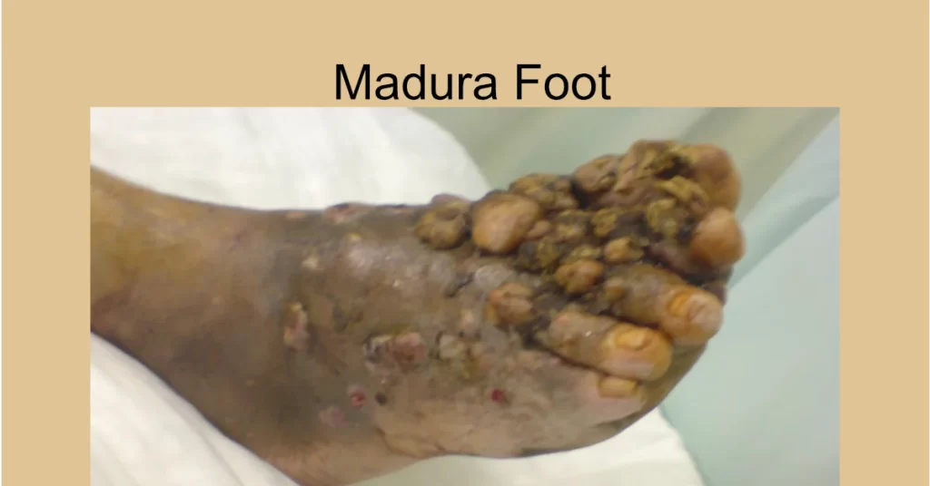
Clinical Features: The clinical features of Madura foot depend on the type of mycetoma. Eumycetoma typically presents as a painless subcutaneous mass with multiple sinuses that discharge a purulent material containing black or white granules. Actinomycetoma usually presents as a subcutaneous mass with multiple sinuses that discharge a purulent material containing yellow or white granules.
- Presentation Following the initial injury, the disease typically follows a slow chronic course over many years with painless swelling and intermittent discharge of pus.
- There may be a deep itching sensation.
- Pain may occur due to secondary bacterial infection or bone invasion.
- After some years, massive swelling of the area occurs, with induration, skin rupture, and sinus tract formation. Lesions contain multiple sinus tracts that usually discharge serosanguinous fluid and at times, grossly visible granules of various colors, sized, and degrees of hardness depending on the agent involved.
- As the infection spreads, old sinuses close and new ones open.
- The exudates are typically granular.
Color of Granules in Madura Foot: The color of the granules in Madura foot is an important diagnostic feature. Black granules are typically seen in eumycetoma, while yellow or white granules are seen in actinomycetoma.
Investigations: The diagnosis of Madura foot is usually made by a combination of clinical examination, imaging studies, and laboratory tests. Imaging studies such as X-rays, CT scans, and MRI can help identify the extent of the disease. Laboratory tests such as histopathology, culture, and molecular testing can help identify the causative agent.
- Microscopy and culture of exudates, and skin biopsy for pathology are necessary to identify the causative organism.
- Serology can be helpful with diagnosis or follow-up care during medical treatment.
- DNA sequencing has been used for identification in difficult cases.
- Plain x-rays are used to assess for evidence of bone involvement.
- CT scan may be more sensitive in the early stages.
- MRI scans can provide a better assessment of the degree of bone and soft tissue involvement; and may be useful in evaluating the differential diagnosis of the swelling.
Differential Diagnosis: The differential diagnosis of Madura foot includes other soft tissue infections such as cellulitis, abscesses, diabetic foot ulcers, chronic bacterial osteomyelitis, tuberculosis, or the early phase of Buruli ulcer.
Management: The management of Madura foot involves a combination of medical and surgical interventions. Antifungal or antibacterial therapy is typically used to treat the infection. Surgical interventions such as debridement, amputation, and reconstructive surgery may also be required.
- Actinomycetomas usually respond better to medical treatment than eumycetomas, which are often difficult to treat. Bone involvement complicates clinical management, often leaving surgical amputation as the only treatment option.
- Due to the slow, relatively pain-free progression of the disease, mycetoma is often at an advanced stage when diagnosed.
- Surgical debridement, followed by prolonged appropriate antibiotic therapy for several months is required for actinomycetoma. Combination therapy with trimethoprim-sulfamethoxazole, dapsone and streptomycin has been used. Rifampin has been used in resistant cases.
- Eumycetomas are only partially responsive to anti-fungal therapy but can be treated by surgery due to their normally well circumscribed nature. Surgery in combination with azole treatment is the recommended regimes for small eumycetoma lesions in the extremities. Madurella mycetomatis may respond to ketoconazole, P. boydii (S. apiospermum) may respond to itraconazole. Other agents of eumycetoma may respond intermittently to itraconazole or amphotericin,B.
Complications: The complications of Madura foot include recurrent infections, chronic pain, disability, and social stigma.
Prognosis: The prognosis of Madura foot depends on the stage of the disease and the extent of the infection. Early diagnosis and treatment can lead to a favorable outcome. However, advanced cases may require extensive surgical interventions and may result in significant morbidity.
In conclusion, Madura foot is a rare and challenging disease that requires a multidisciplinary approach for optimal management. Early diagnosis and treatment can improve the prognosis and prevent significant morbidity.


Responses